Echo planar Imaging (EPI)
| Type of sequence | Philips | Siemens | GE | Hitachi | Toshiba |
| SE - Echo planar | SE-EPI | EPI SE | SE EPI | SE EPI | SE EPI |
| GE - Echo planar | FFE-EPI TFE-EPI | EPI Perf EPIFI | GRE EPI | SG-EPI | FE-EPI |
The echo planar (EPI) is the fastest acquisition method in MRI (100 ms / slice), but with limited spatial resolution. It is based on:
- an excitation pulse, possibly preceded by magnetization preparation
- continuous signal acquisition in the form of a gradient echo train, to acquire total or partial k-space (single shot or segmented acquisition)
- readout and phase-encoding gradients adapted to spatial image encoding, with several possible trajectories to fill k-space (constant [nonblipped] or intermittent [blipped] phase encoding gradient, spiral acquisition etc.).
Preparation and contrast
Contrast in echo planar sequences is determined by excitation pulse and possible magnetization preparation. The various possibilities include:
- GE-EPI: single RF excitation pulse, with no preparation -> T2* weighting
- SE-EPI: pair of 90° - 180° pulses (spin echo type) -> T2 weighting
- IR-EPI: 180° inversion pulse to prepare magnetization then RF excitation pulse -> T1 weighting
- DW-EPI: preparatory pattern for diffusion weighting.
GE-EPI
SE-EPI
DW-EPI
Gradients and k-space filling
To constitute the gradient echo train, a readout gradient is continuously applied, with positive and negative alternations.
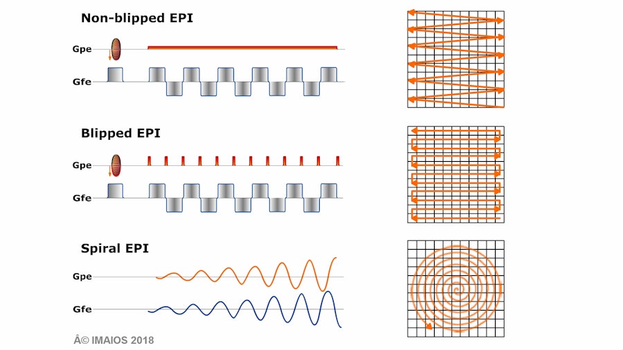
In the case of an alternating gradient (blipped and nonblipped EPI), k-space will be scanned from left to right and back, with each echo. At the same time, the phase encoding gradient may be permanent and constant (nonblipped) giving a zigzag global trajectory, or intermittent (blipped) at each echo onset, giving a rectilinear trajectory.
In the case of spiral k-space filling, phase encoding and readout gradients have a sinusoidal growing envelope.
In all cases, the continuous readout signal imposes k-space regularisation before the image can be reconstructed. The Fourier plane matrix values are calculated by mathematical interpolations that are more or less complex depending on the filling trajectory used.
Echoplanar sequences demand intense, high-performance gradients (for fast signal readout), with short ascent times (because of frequent gradient-switching).
Artifacts
The artifacts in echoplanar sequences are linked to:
- sensitivity to magnetic susceptibility, which can be reduced by using segmented rather than single-shot sequences, at the cost of increasing scan time
- gradient imperfections (particularly induced currents) which perturb spatial encoding, leading to ghost images
- the narrow readout bandwidth in the phase-encoding direction, provoking chemical shift artifacts in this direction, thus requiring suppression of the echoplanar fat signal.
Applications
Echoplanar sequences are the basis for advanced MRI applications such as diffusion, perfusion and functional imagery, to be further dealt with by chapters in the second part.