Magnetic susceptibility and metal
Origin
Magnetic susceptibility corresponds to the internal magnetization of a tissue resulting from the interactions with an external magnetic field. When two tissues with different magnetic susceptibilities are juxtaposed, it causes local distortions in the magnetic field. There are such natural interfaces between air and tissue or between trabecular bone and tissues.
These static field inhomogeneities (T2*) create dephasing and frequency shifts of nearby spins. This results in artifacts in the MR image, mostly a loss of signal, but also a distortion of the image.
The presence of any metal (ferromagnetic or not) causes large distortions in the magnetic field and significant susceptibility artifacts. The range of signal loss depends on the metal and on the pulse sequence (spin echo, gradient echo). The explanations for this signal loss are:
- The local field inhomogeneities (T2*) which accelerate transverse relaxation and signal decay
- The magnetic field distortion so that there is a precessional frequency shift:
- when the slice selection is performed, resulting in an absence of spin excitation and an absence of signal
- when the signal is acquired and the readout gradient is applied, resulting in a shift of spatial localization which causes a signal loss and/or image distortion.
Remedies
Many methods can reduce or modify susceptibility artifacts:
- Spin echo sequences are less prone to susceptibility artifacts than gradient echo sequences. In SE, the 180° refocusing pulse corrects the susceptibility-induced dephasing of spins (which belongs to T2* effects).
- Swapping the frequency-encode and phase-encode directions modifies the susceptibility artifacts without eliminating them.
- Short TE allows less time for dephasing and reduces signal loss.
- A large receiver bandwidth (strong gradients) shortens the minimal TE available.
Usage
Detection of hematomas
Susceptibility artifacts are used to detect hematomas: blood breakdown products (hemosiderin...) cause susceptibility artifacts that are responsible for a signal loss in T2*-weighted GE images.
Quantification of liver iron content
MRI can be useful to quantify the liver iron content. Genetic hemochromatosis causes an iron overload which is stored in the liver. The susceptibility effects of iron account for the decrease in the liver signal intensity, compared to the muscle signal. T2*-weighted gradient echo sequences are the most sensitive for detecting a slight iron overload.
Contrast agents
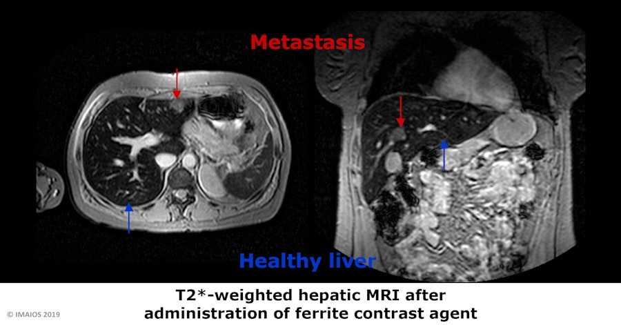
Particulate agents such as ferrites can help to detect liver metastases. They cause a signal loss on T2-weighted images due to magnetic susceptibility effects. These agents are phagocytosed by the reticuloendthelial system of the healthy liver. As a consequence, the normal liver appears dark and the abnormal tissues are highlighted.
Perfusion imaging
Perfusion imaging is based on the modifications of the MR signal when a paramagnetic contrast agent is administered. Adequate pulse sequences (T2* or T2-weighted) analyze the signal loss due to the first-pass of the contrast agent (T2 susceptibility effects of gadolinium). This signal decrease is used to compute the relative perfusion in that region.