
Cholangiopankreatografia rezonansu magnetycznego (MRCP): obrazowanie i anatomia dróg żółciowych, pęcherzyka żółciowego i przewodu trzustkowego
Cholangiopankreatografia rezonansu magnetycznego (MRCP): obrazowanie i anatomia dróg żółciowych, pęcherzyka żółciowego i przewodu trzustkowego
This anatomical module of e-Anatomy is dedicated to the anatomy of the biliary tract on MRI on a MRCP (magnetic resonance cholangiopancreatography).
To emphasize our pedagogic goal in anatomy, we did not choose a healthy patient where the thin (not dilated) biliary tree may have been difficult to see on MRI, but a patient with dilated biliary tree (intrahepatic ducts and common bile duct) on a choledocholithiasis (gallstone) of the distal common bile duct.

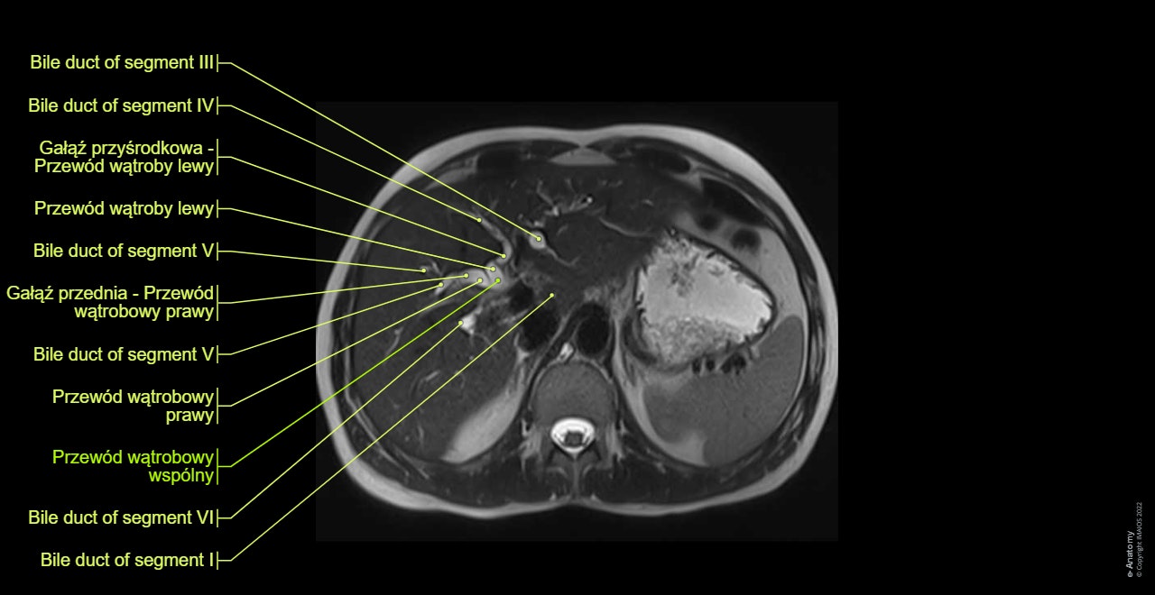
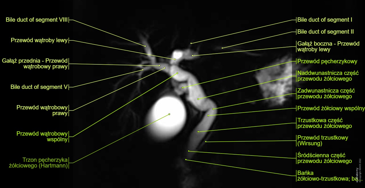
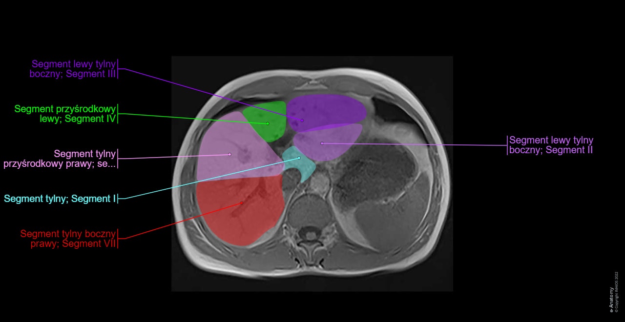
Note that in the case displayed in this module, the patient presents a small anatomical variation of the biliary tree anatomy, type IIIA (the right posterior hepatic duct draining into the left hepatic duct (see diagram below)).
We perform a standard protocol of MRCP (magnetic resonance cholangiopancreatography) on a 1.5 T Siemens Aera:
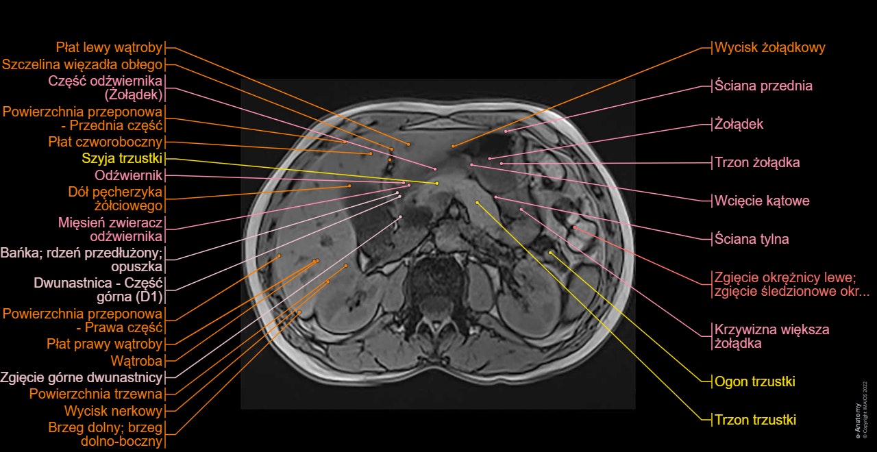
Axial T2 HASTE: Axial breath-hold T2-weighted turbo spin-echo (TSE), 5 mm slices, on the whole biliary tract. Two breath-hold acquisitions are obtained so that the whole of the liver down to the duodenal ampulla is visualized.
MRCP thick slab (T2 weighted breath-hold HASTE fat-saturated thick slab): A fat saturated HASTE sequence where a single slab of data 4 cm in thickness is acquired in a 1- to 2-s breath-hold in 3 different coronal planes. MIP images have also been displayed.
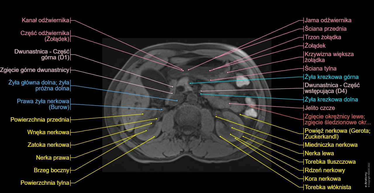
Axial T2 HASTE 4 mm on CBD: An axial breath-hold T2-weighted turbo spin-echo (TSE), 4 mm slices only on the dilated common bile tract.
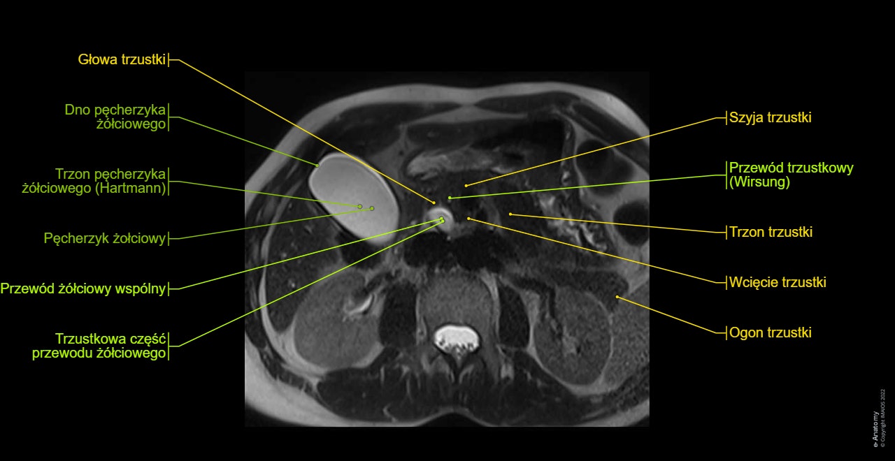
Axial T1 in-phase and axial T1 out-of-phase on the liver: Conventional abdominal MRI imaging to study extraductal structures (hepatic segments, pancreas, spleen, kidneys)
Axial T1 VIBE FS: A 3D fat suppressed T1-weighted GRE sequence without intravenous contrast administration to evaluate the duct walls, and any focal parenchymal pathology.
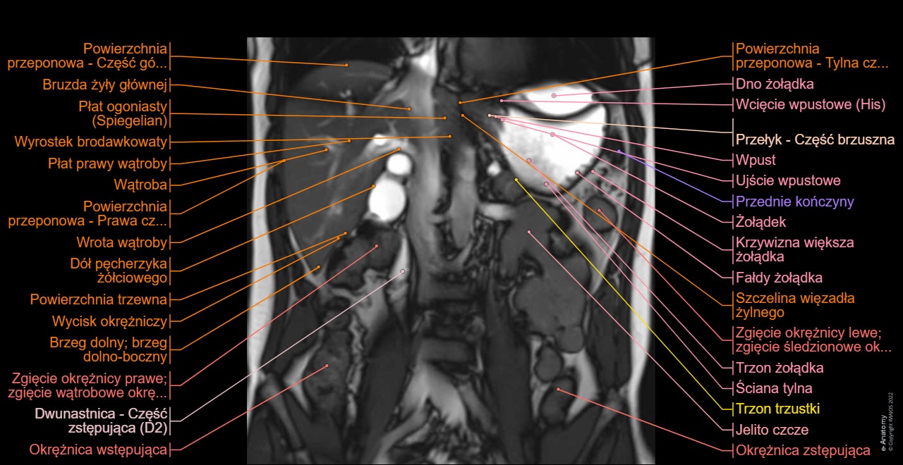
Coronal true FISP: This sequence is the first of our MRCP protocol, on the upper abdomen to establish the location of the extrahepatic bile ducts. But in this module of e-Anatomy, put it at the end because of its lack of relevant medical information on the biliary tract.
- Griffin N, Charles-Edwards G, Grant LA. Magnetic resonance cholangiopancreatography: the ABC of MRCP. Insights Imaging. 2012;3(1):11–21. doi:10.1007/s13244-011-0129-9
- Normal anatomy and anatomic variants of the biliary tree and pancreatic ductal system at MRCP – what the clinicians want to know - J. Ressurreição - ECR 2014 - http://dx.doi.org/10.1594/ecr2014/C-1696
- Terminologia anatomica: international anatomical terminology By the Federative Committee on Anatomical Terminology (FCAT). Stuttgart: Georg Thieme Verlag. ISBN-10: 3-13-114361-4. ISBN-13: 978-3-13-114361-7.